Advances in microscopy over the last century have provided us with an unprecedented view of tiny things in our world that our naked eyes cannot even begin to comprehend. Scanning electron microscopy (SEM), in particular, has given us some striking images over the years to tantalize our visual senses.
The rest of this article is behind a paywall. Please sign in or subscribe to access the full content.SEM involves scanning a focused beam of high-energy electrons over the surface of a sample in order to produce a variety of signals that inform us of certain characteristics of the sample, including topography (surface shape) and chemical composition.
The human eye is able to distinguish two objects 0.2 mm apart; this distance is known as the resolution. Scanning electron microscopes are able to achieve a resolution of under 1 nanometer (10-9 meters) - that’s pretty impressive! They are also able to magnify objects up to around 300,000 times, which is significantly greater than modern light microscopes which reach up to around 1,000 X. Although the images produced are in greyscale they can be colored by software and imaging systems using either true colors or false colors. This is often done merely for aesthetic purposes but it can also help to clarify structure and make the image look more true to life.
Here’s a collection of awesome SEM images of a variety of different things to give you an idea of the incredible detail that SEMs can provide:
Pollen from a variety of common plants including the sunflower, primrose and lily.
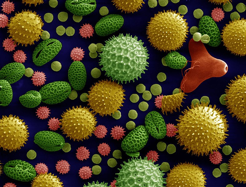
Image credit: Dartmouth Electron Microscope Facility, via Wikimedia Commons.
Trachea epithelium showing ciliated cells (cells with hair-like projections).
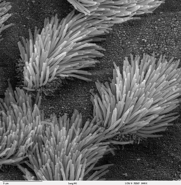
Image credit: Charles Daghlian, Dartmouth Electron Microscope Facility, via Wikimedia Commons.
Head and mouth of a leaf beetle (Chrysomelidae family).

Image credit: Louisa Howard, Dartmouth Electron Microscope Facility, via Wikimedia Commons.
The eye of the fruit fly (Drosophila).
![]()
Image credit: Louisa Howard, Dartmouth Electron Microscope Facility, via Wikimedia Commons.
Yellow mite (Lorriya formosa)

Image credit: Agricultural research service, via Wikimedia Commons.
Bed bug (Cimex lectularius)

Image credit: Centers for Disease control, via Wikimedia commons.
Surface of a kidney stone (ouch...)
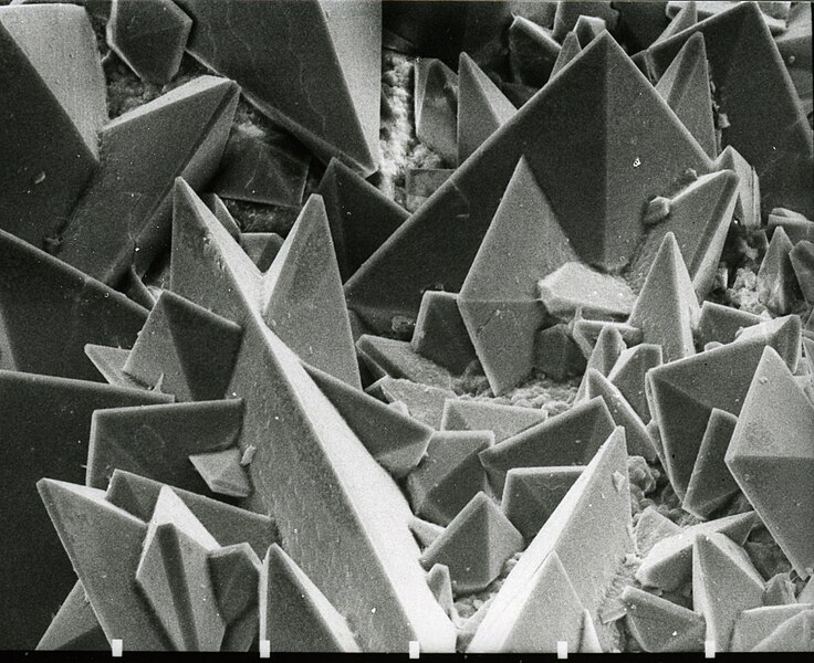
Image credit: Kempf EK, via Wikimedia Commons.
Yersinia pestis, the causative agent of the bubonic plague, on the spines of a flea.

Image credit: NIAID via Wikimedia Commons
White blood cell eating Methicillin Resistant Staphylococcus aureus (MRSA)

Image credit: NIH, via Wikimedia Commons.
Salmonella typhimurium invading human cells
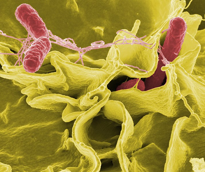
Image credit: Rocky Mountain Laboratories, NIAID, NIH, via Wikimedia Commons.
HeLa cell undergoing programmed cell death (apoptosis).

Image credit: NIH, via Wikimedia Commons
Algae (Gephyrocapsa oceanica)
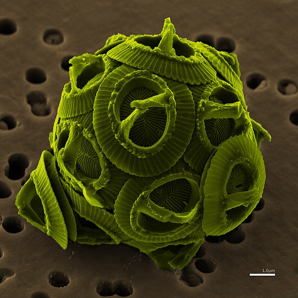
Richard Bartz, via Wikimedia Commons.
Tardigrade (water bear)

Image credit: Goldstein lab, via Wikimedia Commons.