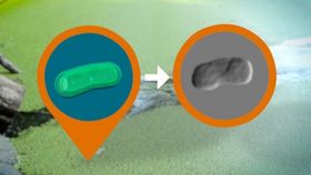


Cyanobacteria, also known as blue-green algae, are small yet mighty organisms. Roughly 2.5 billion years ago, they made the atmosphere on Earth suitable for complex life to breathe and enjoy. A new paper in Nature Communications describes how researchers from SLAC National Accelerator Laboratory were able to take the first X-ray images of these living bacteria; a feat that had previously ended in failure. The research was led by Tomas Ekeberg from Uppsala University in Sweden.
The rest of this article is behind a paywall. Please sign in or subscribe to access the full content.Previously, it had been difficult to complete X-ray imaging of bacteria because the process was too corrosive and the specimens would be obliterated before a clear image could be taken. Using SLAC’s Linac Coherent Light Source (LCLS) X-ray laser, they were able to use shorter pulses, which were able to produce images faster than the radiation could destroy the specimen.
"We have developed a unique way to rapidly explore, sort and analyze samples, with the possibility of reaching higher resolutions than other study methods," one of the paper’s senior authors, Janos Hajdu, said in a press release. "This could eventually be a complete game-changer.”

Image credit: (SLAC National Accelerator Laboratory)
This technique doesn’t require specimens to be pre-treated. With no dyes or genetically-engineered biomarkers required, the microbes can be viewed in their natural, unadulterated state. The information gathered from this form of imagery has plenty to teach researchers about the shape and internal structure of bacteria and viruses, which will have a wide variety of biomedical applications.
"You can study the full cycle of cellular processes, with each X-ray pulse providing a snapshot of the process you want to study,” Ekeberg explained.
SLAC’s LCLS works by spraying the specimens into the air and then pulsing the X-rays rapidly. This technique is able to obtain 100 images per second, which means that a great deal of useful data can be collected in a fairly short order. Rather than just having a single snapshot of the specimen, the researchers will have a wealth of images to consider and really get an accurate depiction of the processes in chronological order.
"One can start to analyze differences and similarities between groups of cellular structures and show how these structures interact: What is in the cell? How is it organized? Who is talking to whom?” continued Hajdu.
Moving forward, researchers hope to further develop the technology and improve the resolution in order to better see the internal structures of these microbes.