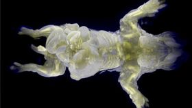


One of the limitations of studying very fine and delicate cellular connections is that it is hard to see exactly what’s going on because of the color of the cells. Clearing the body of heme, which is part of hemoglobin, has allowed scientists from the RIKEN Quantitative Biology Center in Japan to create mice that are almost completely transparent. This will allow structures to be imaged in unprecedented detail in order to better understand their function. Kazuki Tainaka and Shimpei I. Kubota were co-lead authors of the paper that was published in Cell.
The rest of this article is behind a paywall. Please sign in or subscribe to access the full content.This is not the first attempt at obtaining clear organs. A team from Stanford uses a technique called CLARITY to make brains transparent for imaging purposes. One of the researchers from that team went on to lead a group that applied a similar technique to an entire mouse body. Though many of the structures became transparent, they weren’t able to completely eradicate the heme from the bodies.
The RIKEN group used a similar process called Clear, Unobstructed Brain Imaging Cocktails and Computational Analysis (CUBIC) that was better equipped to not only remove heme from the brain, but also from the heart, kidneys, livers, and lungs. CUBIC worked equally well on both young and old mice.
Credit: RIKEN
"We were very surprised that the entire body of infant and adult mice could be made nearly transparent by a direct transcardial CUBIC perfusion coupled with a two-week clearing protocol. It allowed us to see cellular networks inside tissues, which is one of the fundamental challenges in biology and medicine,” Tainaka said in a press release.
The process of clearing out the heme is used on mice that are already dead, and takes about two weeks. Once the specimen is transparent, the scientists can collect 3D images of tissues and organs through the use of light-sheet fluorescent microscopy. The team was already able to see differences in the pancreases of diabetic and non-diabetic mice. The isles of Langerhans, which produce insulin, had clear morphological differences when observed unfettered. Discoveries such as these could eventually lead to new treatments.
"This new method could be used for 3D pathology, anatomical studies, and immunohistochemistry of entire organisms,” added senior author, Hiroki Ueda. “For example, it could be used to study how embryos develop or how cancer and autoimmune diseases develop at the cellular level, leading to a deeper understanding of such diseases and perhaps to new therapeutic strategies. It could lead to the achievement of one of our great dreams, organism-level systems biology based on whole-body imaging at single-cell resolution.”
Image credit: Tainaka et al., 2014
[Hat tip: HuffPo]