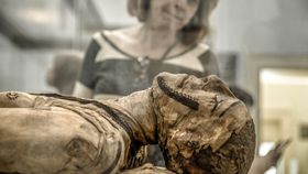


Archaeologists have dabbled with using X-rays to study ancient mummies since the turn of the 20th century. Some 120 years on, the imaging techniques used to learn about the life and death of these people have become unbelievably powerful. Researchers in Sweden have developed a technique that can image the soft tissue of an ancient Egyptian mummy down to a microscopic level – even picking up on the remains of individual cells.
As reported in the journal Radiology, researchers from KTH Royal Institute of Technology/Albanova University Center in Stockholm, Sweden have used a computed tomography (CT) technique to image the soft tissue of an ancient Egyptian mummy's hand. Within these images, we are able to see the finest biological details of the mummy, with a resolution between 6 and 9 micrometers – that’s just a little bigger than a human red blood cell.
The hand was brought from Egypt to Sweden at the end of the 19th century and dates back to around 400 BCE. The revolutionary new technique even allowed the researchers to see nerves, the blood vessels in the nail beds, different layers of the skin, and even the remains of adipose (fat-storing) cells.

Not only will this technique be used to create some ultra-high definition images to document ancient mummies, but it could also even be used to increase our understanding of ancient diseases.
"For studying bone and other hard, dense materials, absorption contrast works well, but for soft tissues, the absorption contrast is too low to provide detailed information," Jenny Romell, from KTH Royal Institute of Technology/Albanova University Center, said in a statement.
"This is why we instead propose propagation-based phase-contrast imaging."

Propagation-based phase-contrast imaging detects both the absorption and phase shift that occurs as X-rays pass through a sample. This creates different degrees of contrast within an image and allows for subtler details to emerge, not just a simple black-and-white projection.
"There is a risk of missing traces of diseases only preserved within the soft tissue if only absorption-contrast imaging is used," Romell added.
"With phase-contrast imaging, however, the soft tissue structures can be imaged down to cellular resolution, which opens up the opportunity for detailed analysis of the soft tissues."

It's still relatively early days for the technique, especially in this field, but the researchers argue that it could someday be widely used in the study of mummies and other ancient remains.
"Just as conventional CT has become a standard procedure in the investigation of mummies and other ancient remains, we see phase-contrast CT as a natural complement to the existing methods," Romell said.