

Scientists can grow some pretty amazing things in the lab these days. From penises to burgers, spinal cords to mini brains, tissue culture has been improving exponentially in recent years. Now, scientists have created a system that allows them to successfully grow tiny beating “hearts” from stem cells, complete with microscopic chambers.
Although this isn’t the first time that miniaturized hearts have been grown in a lab, this newly developed method is an improvement on earlier techniques due to the addition of physical cues that help prompt and guide cell fate during the differentiation process, meaning structure and cell organization can be controlled to a greater extent. This template, the researchers say, could help in the search for new drugs or the testing of existing drugs, and will hopefully further our knowledge of how the heart develops.
“We believe it is the first example illustrating the process of a developing human heart chamber in vitro,” University of California Berkeley professor and co-senior author Kevin Healy said in a statement. “This technology could help us quickly screen for drugs likely to generate cardiac birth defects, and guide decisions about which drugs are dangerous during pregnancy.”
To create these teeny hearts, researchers from UC Berkeley and the Gladstone Institute of Cardiovascular Disease started off by engraving circular patterns into the surface of tissue culture dishes, which were designed to act as a physical guide for cell growth. They then seeded the dishes with a type of stem cell known as human induced pluripotent stem cells, which are skin cells that have been genetically reprogrammed to a blank slate state. By adding a cocktail of chemical cues, the researchers were able to prompt them to differentiate into different cardiac cell types, including heart muscle cells, or cardiomyocytes.
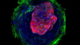
After around two weeks of growth, the scientists observed that the cells had grown and developed from 2D monolayers to the beginnings of 3D structures, complete with beating microchambers. Importantly, they self-organized in such a manner that the central region mainly consisted of muscle cells, whereas those on the periphery were mostly the type of cells that form connective tissue, or fibroblasts. And thanks to the patterned templates, each colony of cells was of uniform size and shape. These findings have been published in Nature Communications.
“We envision these micro hearts recapitulate an early stage of human heart development, which can be considered as a checkpoint for a healthy fetus” lead author Zhen Ma told IFLScience. “We can use these micro hearts to screen drugs, which might lead to malformation of the embryonic heart.”
For example, the researchers exposed the differentiating stem cells to a drug called thalidomide, a sedative that can cause severe birth defects and alter embryonic heart development. As anticipated, the microchambers failed to develop normally when compared with controls, demonstrating reduced muscle contraction and beat rates.
This supports their proposed use for drug testing, which would be a particularly useful application. Each year, co-senior author Dr. Bruce Conklin explains, hundreds of thousands of pregnant women are given drugs that could potentially harm the developing fetus. Hopefully, this system could ultimately help reduce this risk.