


A breakthrough imaging technique provides spectacular flythrough views of the brain's inner connections -- intricate structures that are typically lost when the brain is cut up into thin slices. A new suite of improvements was published this month, and the latest rendition could help the technique go mainstream.
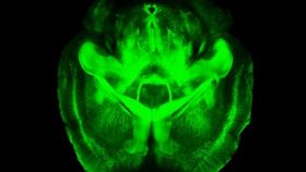
Last year, Stanford's Karl Deisseroth and colleagues revealed a new way to peer into an intact brain through a process that renders the whole organ transparent. Existing methods typically require that the brain be sliced, which compromises the ability to analyze how components relate to each other. With their technique -- called CLARITY -- the postmortem brain isn't sliced or sectioned in any way, which allows the three-dimensional complexity of its wiring and molecular structures to be measured and probed.

To do so, the researchers extracted the opaque elements -- lipids, in particular -- from the brain while keeping the important features unscathed. Those fatty molecules help much of the brain's structure, so to keep the remaining brain tissue from falling apart without them, the team replaced the brain's lipids with a water-based gel. The brain is immersed in the hydrogel solution so individual molecules called hydrogel monomers can infuse the tissue. When heated, the monomers congeal into polymer chains that form a mesh throughout the brain. This mesh holds it all together while the team extracts the lipids (which have been dissolved in the electrically-charged detergent) using an electric field. What remains is a 3D, transparent brain with all its neurons, axons, dendrites, synapses, proteins, and nucleic acids in place. Pictured here, an intact mouse brain before and after the 2-day CLARITY process.
With no more fat to block the light, the team was able to get visible light and macromolecular probes to penetrate the tissue and conduct a 3D molecular analysis of the intact brain. Fluorescent antibodies can help light up specific structures and literally illuminate the chemical relationships of cells and subcellular structures in the brain.
Below are their videos of a rodent brain. The first is a flythrough of an intact mouse brain, and the second is a 3D view of a mouse brain’s memory hub, or hippocampus, comprised of neurons (green), interneurons (red), and support cells (blue).
However, even though it has since been used by labs around the world, the system hasn't been broadly adopted. To make CLARIFY safer and more reliable, the latest version improves on the original with technological fixes for two distinct challenges.
First, the electric field aspect was challenging for some labs; the voltage sometimes damaged the tissue. So Deisseroth's team devised an alternative way of pulling fat from the hydrogel-embedded brain. It takes a little longer, but still removes all the fat and requires nothing more than some chemicals and a warm bath.
Second, the probes stopped working when they've been exposed to too much light -- a problem for time-consuming, high-resolution images of whole brains. So the team made it possible to scan an entire plane at one time, instead of a point, which buys a couple orders of magnitude of time.
Their paper is the first to be published with support of the White House BRAIN Initiative, which seeks to map the brain to help us understand how we think, learn, and remember. As CLARITY becomes more widely used, the team hopes it will continue to reveal how inner circuits are structured in normal and diseased brains, pointing the way to new therapies.
The work was published in Nature Protocols last week.
[Via Stanford]
Image: Deisseroth lab