


Like one of Frankenstein's lesser-known side projects, neuroscientists and mechanical engineers at the University of Minnesota have transplanted see-through implants into the tops of mouse skulls, allowing them to watch the drama of the brain unfold in front of their eyes.
With new insights already coming out of the research, the team hopes their implants could be used to get an unprecedented glimpse into the nature of the human brain and conditions such as concussions, dementia, Alzheimer’s disease, and Parkinson’s.
“What we are trying to do is to see if we can visualize and interact with large parts of the mouse brain surface, called the cortex, over long periods of time. This will give us new information about how the human brain works,” co-author Suhasa Kodandaramaiah, PhD, a mechanical engineer at the University of Minnesota, said in a statement.
“This technology allows us to see most of the cortex in action with unprecedented control and precision while stimulating certain parts of the brain.”
As you can see in the video below, the window into the brain and mesoscale imaging allow scientists to watch as the brain's cortical surface flashes with activity.
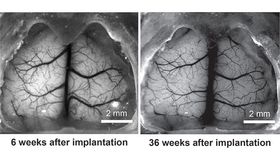
Reporting in the journal Nature Communications, the team created the “See-Shell” transplant using digital scans of the mouse skull and 3D-printing technology. They then surgically transplanted the transparent polymer skull tops into dozens of mice for an average of 92 days, although the implant can remain in place for over 300 days.
Over 90 percent of the transplants were a success and the body did not reject the implant, allowing the researchers to study the same mouse brain over a period of several months. This, the researchers argue, opens the possibility of studying the effects of chronic conditions, degenerative diseases, and aging on the brain.
The first study using the See-Shell device looked to see how a mild concussion in one part of the brain can affect other parts of the organ. Using this window into the brain, the researchers learned how the brain reorganizes itself, both structurally and functionally, following the physical trauma.
“This new device allows us to look at the brain activity at the smallest level zooming in on specific neurons while getting a big picture view of a large part of the brain surface over time,” Kodandaramaiah added. “Developing the device and showing that it works is just the beginning of what we will be able to do to advance brain research.”