


Researchers in Japan have developed a new system for keeping tissues alive outside the body, and have used it to preserve the functionality of slices of mouse brains in a lab for 25 days. The team behind this development are hopeful that their work will facilitate the creation of countless new medications by allowing pharmacologists to test drugs on live tissue for extended periods of time.
At present, it is extremely difficult to keep complicated tissue alive for more than a few days once it has been removed from a body, which limits opportunities for testing new drugs. This is because tissues dry out very quickly, so need to be kept in a liquid medium that contains the appropriate nutrients, although this tends to "drown" the tissue, preventing it from respiring by blocking the transfer of gases between cells.
To overcome this hurdle, scientists at the RIKEN Center for Biosystems Dynamics Research developed a microfluidic device, which keeps tissue in constant contact with a liquid medium without immersing it, thus ensuring it neither dries out nor drowns.
The device consists of a semi-permeable channel that is surrounded by an artificial membrane and solid walls made from a compound called polydimethylsiloxane (PDMS), which is used as an anti-foaming agent in many common medications.
A culture medium containing the right nutrients is allowed to circulate through the channel, nourishing the tissue through the membrane while simultaneously removing waste products from its biological processes.
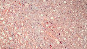
Describing their work in the journal Analytical Sciences, the researchers explain how they tested their device using slices of tissue from a part of the mouse brain called the suprachiasmatic nucleus, which regulates the rodents’ circadian rhythms, or body clock. First, though, they had to create a strain of genetically engineered mice that produced fluorescent proteins to help regulate their circadian rhythms.
By tracking the production of these brightly colored proteins, the team was able to measure the functionality of the suprachiasmatic nuclei of these mice once they had been extracted from the animals’ heads.
After a little trial and error regarding the rate at which the culture medium was allowed to flow through the semi-permeable channel, the team were able to keep these brain slices alive and functioning for 25 days, at which point they stopped the experiment.
Using conventional methods, brain tissue can only be kept alive for a few days in a laboratory, and a drop in neural activity of 6 percent is seen in the first 10 hours. Yet the microfluidic device enabled neural activity to remain at 97 percent after the 25 days had elapsed, prompting the study authors to suggest that their method could be used to keep brain tissues viable for over a hundred days.
Looking at the potential future applications of this system, study co-author Nobutoshi Ota said in a statement that it could be used to “improve research into organogenesis through long-term culturing and observation which is necessary for growing tissue and organs.”