


A tiny Egyptian sarcophagus that was long believed to hold the remains of a mummified hawk actually tells of one family's tragic story. Recent micro-CT scans have determined that the contents of the 2,100-year-old tomb contain the remains of a near-to-term stillborn male fetus with severe congenital abnormalities, including a malformed skull and vertebrae.
An international team of more than a dozen experts “virtually unwrapped” the mummy to reveal what they say would have been a “family tragedy even two millennia ago.” Hidden behind the ancient layers is a male fetus, probably between 23 and 28 weeks of gestation.
"The whole top part of his skull isn't formed. The arches of the vertebrae of his spine haven't closed. His ear bones are at the back of his head," said bioarcheologist Andrew Nelson, who led the examination, in a statement.
The researchers believe the fetus suffered from a rare condition called anencephaly, a serious birth defect in which a baby is born without parts of the brain and skull – a condition that today affects about less than 1 percent of births worldwide. According to the researchers, it is just one of two anencephalic mummies known to exist and is the most-studied fetal mummy in history.

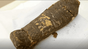
Micro-CT scans determined that a "hawk mummy'" at Maidstone Museum UK is, in fact, a stillborn male human with severe congenital abnormalities, including a malformed skull and vertebrae. The scans are the most detailed of any fetal mummy yet. Maidstone Museum UK/Nikon Metrology UK
"It would have been a tragic moment for the family to lose their infant and to give birth to a very strange-looking fetus, not a normal-looking fetus at all,” said Nelson, who continues that this fetus could be special in providing important clues to how people lived at the time. Anencephaly can result when the mother’s diet lacks folic acid found in green vegetables, which could lend more information as to how Egyptians ate at the time. It also raises new questions about what circumstances led to mummification. It was previously thought fetuses were only mummified when they were believed to have some power as talismans.
The mummy’s misidentification was revealed two years ago when the Maidstone Museum CT-scanned its resident female mummy and other animal mummies at the time. Scans of “EA 493 – Mummified Hawk Ptolemaic Period” surprised mummy experts when they showed a mummy that resembled that of a human.
Nelson then conducted a micro-CT scan – a high-resolution scan that didn’t damage the mummy – and consulted a team of specialists in Egyptology, radiology, anatomy, neonatology, and urology to interpret the scans. They saw well-formed toes and fingers, but no bones to shape the roof and sides of the skull where the brain would normally grow.
“In this individual, this part of the vault never formed and there probably was no real brain,” Nelson said. He presented the team’s findings at the Extraordinary World Congress on Mummy Studies in the Canary Islands.