


Researchers seeking to unlock the secrets of how the brain works have created the largest and most detailed map of connections between brain cells ever produced. Though the resulting images may look like a Jackson Pollock painting, they actually reveal the neurological activity of a live mouse as it responds to visual stimuli, providing key insights into the ways in which different brain cells interact with one another as they encode information.
The work – which appears in the journal Nature – was conducted as part of an attempt to build on previous studies into brain connectivity and answer some of the major unresolved questions regarding this topic. For instance, prior research has indicated that neurons that respond to highly similar visual stimuli tend to form stronger connections to one another than neurons responsible for unrelated processes.
However, scientists are unsure whether this occurs because neurons with a similar orientation form larger connections – known as synapses – or because they simply form a greater number of synapses. Alternatively, it has been proposed that this type of selective connectivity may be regulated by the spatial arrangement of synapses, which may occur in clusters where these neurons meet, thereby producing a strengthened point of connection.
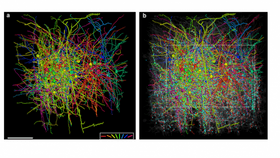
To investigate, researchers used a custom-designed two-photon laser-scanning microscope to create a detailed image of a small section of the visual cortex of a live mouse as it watched a screen displaying moving horizontal and vertical lines. The visual cortex is the part of the brain responsible for processing visual stimuli, with different but highly connected neurons responding to the different types of lines.
Not only did this reveal a physical picture of the arrangement of neurons in this part of the brain, but by tracking the changes in calcium concentrations within neurons, the researchers were able to ascertain patterns of activity.
Following this, the team removed the relevant section of the mouse’s brain and used a technique called electron microscopy to complete the full picture of connections between neurons. In total, they found that the tiny piece of brain under observation contained 201 neurons, which formed a total of 1,278 synapses with one another.

Brain cells – or neurons – connect at junctions called synapses. nobeastsofierce/Shutterstock
As expected, connectivity was much stronger between neurons that conducted similar processes than between those that did not, despite the fact that these cells were not arranged in any particular spatial order that might produce this phenomenon. Indeed, the neuronal spines – known as axons and dendrites – of these particular cells passed by one another with the same frequency as those of less related neurons, yet when they did so they tended to form more synapses.
These synapses were also larger than those occurring between neurons that perform unrelated functions, providing a greater surface for the transmission of chemical messengers called neurotransmitters, resulting in a stronger signal. However, these synapses were not arranged in clusters, as had been hypothesized.
While this work still leaves us with a long way to go in order to fully understand the ways in which the electronic highways crisscrossing the brain combine to generate consciousness, it does represent a first step on the road to building an accurate map of how these connections occur. Signing off, the study authors urge future researchers to continue this project, insisting that the perfection of this process will lead to “a richer understanding of [the brain’s] network structure and function.”