


Some cancers appear indistinguishable from healthy tissue, raising the risk of recurrence and metastasis. Now, researchers in Canada have developed a handheld fiber optic probe that they say can detect invasive brain cancer in patients accurately. The device, described in Science Translational Medicine this week, could one day quickly guide neurosurgeons during procedures to remove cancerous tissue from the brain.
Glioma is a type tumor that forms from the glial cells that surround and support nerve cells in the brain and spinal cord. Diffusely invasive gliomas are aggressive and can infiltrate healthy tissue extensively. “Often it is impossible to visually distinguish cancer from normal brain, so invasive brain cancer cells frequently remain after surgery, leading to cancer recurrence and a worse prognosis,” McGill University’s Kevin Petrecca says in a news release. “Surgically minimizing the number of cancer cells improves patient outcomes.”
So, Petrecca and colleagues developed a probe based on Raman spectroscopy, a technique utilizing lasers to measure the way molecules in an object scatter light, producing a spectrum that’s unique to that object. “The emitted light provides a spectroscopic signal that can be interpreted to provide specific information about the molecular makeup of the interrogated tissue,” says study author Frederic Leblond of Polytechnique Montréal. Once in contact with the brain, the probe illuminates a small spot about a millimeter deep into the tissue and provides real-time Raman spectra of that area.
 The team tested their probe (pictured to the right) with 17 neurosurgical patients who have advanced gliomas. In addition to dense tumor masses, the device was able to detect individual cancer cells that have invaded the surrounding tissue with 92 percent accuracy.
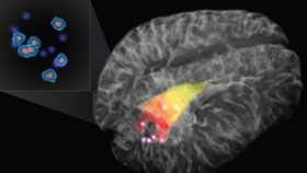
In the 3D rendering above, the red and yellow areas indicate cancer that was detected using an MRI. The bright points—cancers detected using Raman spectroscopy—are well beyond what’s detectable using MRI. The Montreal Neurological Institute and Hospital will be launching a clinical trial for patients with newly diagnosed and recurrent glioblastoma.
Images: Laboratory for Radiological Optics/Polytechnique Montreal and Montreal Neurological Institute (top), McGill University (middle)