Advertisement

clockPUBLISHED

Mind-Blowing Images From This Year's Nikon Photomicrography Competition

author

Tom Hale

author

Tom Hale

Senior Journalist

Tom is a writer in London with a Master's degree in Journalism whose editorial work covers anything from health and the environment to technology and archaeology.

Senior Journalist

comments1Comment

1st place winner: Yousef Al Habshi, United Arab Emirates. Eye of a Metapocyrtus subquadrulifer beetle Reflected Light 20x (objective lens magnification). Yousef Al Habshi/Nikon Small World Photomicrography Competition 

Feast your eyes on the winners of the 2018 Nikon Small World Photomicrography Competition, an annual photography competition dedicated to finding the beauty and scientific intrigue in all things stupidly small, from spider embryos to human teardrops.

Emirati photographer Yousef Al Habshi is this year’s winner for his perspective-bending image of a Metapocyrtus subquadrulifer Weevil. Although this species of beetle is typically smaller than 11 millimeters (0.4 inches), Al Habshi’s image clearly picks up on the insect’s compound eye and surrounding greenish scales with unbelievable clarity.

Advertisement

“Because of the variety of coloring and the lines that display in the eyes of insects, I feel like I’m photographing a collection of jewelry,” Al Habshi said in a statement. “Not all people appreciate small species, particularly insects. Through photomicrography, we can find a whole new beautiful world which hasn’t been seen before. It’s like discovering what lies under the ocean’s surface.”

His image is much more than a pretty picture, too. Al Habshi’s work has been used by Professor Claude Desplan, of New York University Abu Dhabi, to help understand the lifecycle of M. subquadrulifer weevils and how farmers can ward off infestations.

2nd Place: Rogelio Moreno, Panama. Fern sorus (structures producing and containing spores). Autofluorescence 10x. Rogelio Moreno/Nikon Small World Photomicrography Competition  

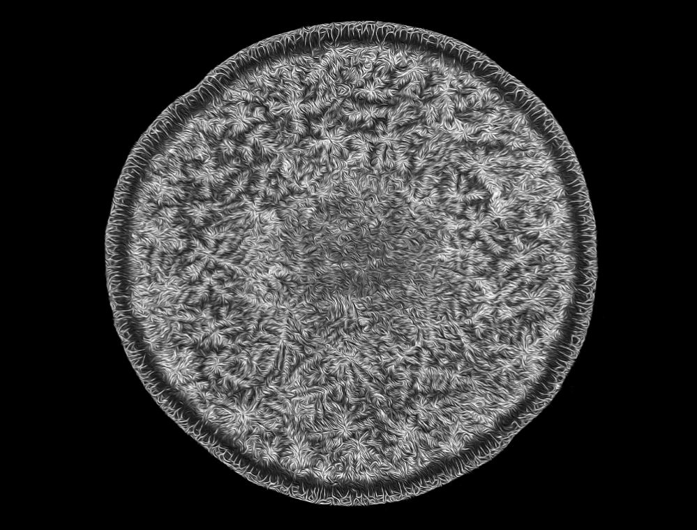
Second place was awarded to Rogelio Moreno from Panama for his colorful photo of a fern’s sorus, the microscopic structures found in ferns (and fungi) that produce spores, using a photo stacking technique.

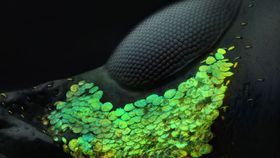
Saulius Gugis of Illinois in the US took home third place for his image of a spittlebug nymph. The tiny spittlebug was captured in the middle of making a “bubble house”, a frothy layer of foam it creates to hide from predators, keep warm, and stay moist.

Advertisement

You can check out some of the other winning images, along with the “honorable mentions” from the judges, below. If this kind of stuff floats your boat then be sure to check out previous years' entries.

3rd Place: Saulius Gugis, Illinois, USA. Spittlebug nymph in its bubble house. Focus Stacking 5x. Saulius Gugis/Nikon Small World Photomicrography Competition  
4th Place: Can Tunçer, Turkey. Peacock feather section. Focus Stacking 5x.Can Tunçer/Nikon Small World Photomicrography Competition
5th Place: Dr Tessa Montague, Massachusetts, USA. Parasteatoda tepidariorum (spider embryo) stained for embryo surface (pink), nuclei (blue), and microtubules (green). Confocal 20x. Dr Tessa Montague/Nikon Small World Photomicrography Competition
7th Place: Norm Barker, Maryland, USA. Human tear drop. Darkfield 5x. Norm Barker/Nikon Small World Photomicrography Competition
8th Place: Pia Scanlon, Australia. Portrait of Sternochetus mangiferae (mango seed weevil). Stereomicroscopy, Image Stacking 1x. Pia Scanlon/Nikon Small World Photomicrography Competition
19th Place: Pierre Anquet, France. Asian hornet (Vespa velutina) with venom on its stinger. Reflected Light, Focus Stacking 6.3x. Pierre Anquet/Nikon Small World Photomicrography Competition

ARTICLE POSTED IN

  • tag
  • photography,

  • microscope,

  • art,

  • insect,

  • culture,

  • photo

FOLLOW ONNEWSGoogele News