


Our understanding of the behavior and structure of DNA has just taken a massive leap forwards, thanks to new high-resolution images that reveal how the molecule writhes, wriggles and squirms in order to squeeze itself into cell nuclei. Appearing in the journal Nature Communications, the incredible new images allowed researchers to observe individual atoms within a DNA molecule, tracking their movement as they perform their contortionist dance.
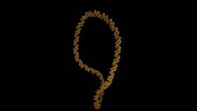
Most people are familiar with the iconic double-helix structure of DNA, yet this neat, static image doesn’t accurately reflect the complex shapes that the molecule adopts in real life. In order to cram itself into the nucleus of a cell, our genetic material must twist and coil around itself, a process known as supercoiling. Doing so allows for a staggering two meters (6.6 feet) of DNA to pack itself into every cell in the human body.
The new images reveal that DNA is much more dynamic than previously thought, and is constantly dancing in order to form an ever-changing array of diverse shapes. According to the study authors, the more unusual shapes a molecule adopts, the more opportunities it has to bind with other substrates, thereby increasing its capacity to carry out its various functions.
Reacting to these amazing images, study author Dr Alice Pyne from the University of Sheffield commented that “seeing is believing, but with something as small as DNA, seeing the helical structure of the entire DNA molecule was extremely challenging.”
“The videos we have developed enable us to observe DNA twisting in a level of detail that has never been seen before.”
To produce the images, the team used a combination of atomic force microscopy and molecular dynamics simulations. While previous microscopic techniques have only managed to generate static images of DNA, this new approach allowed the researchers to create dynamic sequences that reveal the molecule’s twisting motions.
For this study, the authors looked specifically at DNA minicircles, which are joined at the ends in order to form a loop. The samples they observed were engineered and isolated from bacteria, but have previously been found to occur naturally in cells.

When they examined these minicircles in their relaxed, uncoiled state, the researchers noted that the DNA remained rather motionless. However, once they twisted this genetic material, they found that it came to life and began jiggling around in order to adopt a succession of exotic poses.
Baylor College of Medicine biologist Lynn Zechiedrich, who made the minicircles for the study, commented that the images indicate just how “wrinkled, bubbled, kinked, denatured, and strangely shaped” DNA becomes when it is squeezed into a cell.

According to the researchers, increasing our understanding of the ways in which DNA folds itself into compact shapes could be the key to developing new genetic therapies for a range of untreatable medical conditions.