


Olympus has revealed the winners of its first-ever Global Image of the Year Life Science Light Microscopy Award photography competition, a contest to celebrate and recognize both the scientific and artistic value of life science images across the world. These incredible images reveal the detail, and surprising beauty, in the microcosmos around and inside us.
The rest of this article is behind a paywall. Please sign in or subscribe to access the full content.Starting off life in 2017 as the Olympus Image of the Year European Life Science Light Microscopy Award, the now global competition has four winners, one for each geographical macro-area, and an overall winner.
The global winner crown goes to Ainara Pintor, a graduate researcher in Spain, with her beautiful image (below) of a mouse’s hippocampus, the part of the brain that plays a crucial role in the consolidation of long- and short-term memories.
“I jumped when I realized that my image was selected to be the 2019 Olympus Image of the Year,” Pintor Rial told IFLScience. “It is a fantastic feeling that this image will cross borders, and it will be seen all over the world!”
“I took this picture in October when I was starting to perform immunofluorescence in brain slices. I am looking into the localization of proteins playing an essential role in memory formation, such as Fat mass and obesity-associated (FTO) protein. It is incredible how this fixed tissue seems to be so alive. One word came to my mind: ‘Neurogarden.’ The brain complexity is just unbelievable!”
The four winners were selected from over 400 submissions from 65 countries around the world. The judges evaluated the images on artistic and visual aspects, as well as their scientific impact and the photographer’s microscope proficiency.
“I’m so impressed by the amazing response to our first Global Image of the Year Award,” said Satoshi Nakamura, the Vice President of Scientific Solutions Global Marketing for Olympus. “The creative image submissions embody our contest’s mission of celebrating art in science. We hope this competition continues to inspire people to find beauty in an unexpected place—right under their microscope.”

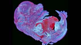
The three regional winners have also delivered gorgeous visuals of life under a microscope.
The Asia winner, Howard Vindin of Australia, captured the autofluorescence of a mouse embryo, with 950 tiles stitched together to create this striking photo.

More mouse highlights were revealed, this time in a frozen section of a mouse head taken by the UK's Dr Alan Prescott, who won the Europe, Middle-East, and Africa title with his photograph "The Mouse's Whiskers".
"This was a part of a section not relevant to the research I was involved in-mainly looking at mitophagy (mitochondrial autophagy) in the brain and eye. The developing whiskers caught my eye, whilst I was scanning the sections, as an aesthetically interesting region. So I took a slightly higher resolution image with a view to using it for a competition or outreach event," Dr Prescott told IFLScience.

The winner for the Americas, Professor Tagide de Carvalho, captured a beautiful image of a tardigrade, incredibly tiny but tough little critters that can survive under extreme conditions.

Nine Honorable Mentions were also selected, with images ranging from flower buds to the wings of insects and a mouse's spinal cord. You can check them all out below as well on the Olympus Life Science website. Who says science can't be beautiful?