


While it is still not entirely clear how shock waves produced by explosions harm those who are exposed to them, a growing body of evidence suggests that they may cause tiny bubbles to form and then explode inside the brain, potentially causing a range of serious injuries.
The rest of this article is behind a paywall. Please sign in or subscribe to access the full content.According to Timothy Bentley of the U.S. Office of Naval Research (ONR), these shock waves travel “faster than the speed of sound,” producing a level of energy that “can cause subtle yet damaging [health] effects,” which even the most advanced military body armor is powerless to prevent. To find out how this occurs, the ONR has been supporting the research of Michael Cho from the University of Texas, whose team has conducted a number of tests into the effects of shock waves on brain tissue.
In a recent paper that appeared in the Journal of Neurotrauma, the researchers explained how several previous studies have shown that shock waves cause tiny bubbles – called microcavitations – to form in water. Because it is not possible to observe the biological impact of shock waves when soldiers are in action, it is not known if this same effect occurs in the brain when explosions occur on the battlefield.
However, Cho and his team suspect that these microcavitations may form in the cerebrospinal fluid of military personnel exposed to blasts, and designed an experiment to examine what effect this might have on brain cells. To do so, they created tissue models using brain cells called astrocytes, which were placed in a fluid through which electrical charges were passed in order to generate shock waves.
Astrocytes make up around 20 to 50 percent of the volume of human brains, and outnumber neurons by about 10 to 1. They are involved in a number of vital processes associated with protecting the central nervous system from damage.

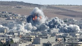
A condition called blast traumatic brain injury has been associated with post-traumatic stress disorder, although the causative link between the two has eluded scientists. John Gomez/Shutterstock
After creating the shock waves, the researchers noted that microcavitations appeared in the fluid around the cells, before bursting after a maximum of 2 seconds. Using a range of chemical markers, they were then able to track the processes occurring in these cells once these tiny bubbles had disappeared, and found that exposure to these microcavitations had a significant negative impact on cell function.
For instance, they found an increase in concentrations of a compound called superoxide, which is known to play a role in apoptosis – or cell death – via a process known as oxidative stress. This occurs when highly reactive oxygen species damage the fabric of a cell. Additionally, the amount of material able to penetrate into cell nuclei increased after microcavitation, indicating that the membrane around the nucleus had become damaged.
Interestingly, those cells that were exposed to electrical charges but not microcavitations displayed none of these effects, suggesting that the bubbles themselves were indeed the cause of this damage. Exactly how this occurs is still not entirely clear, although the study authors suggest that it could be generated by an increase in temperature within these bubbles, or by the release of a powerful ‘water jet’ as they explode.
Effects such as these could be responsible for the condition known as blast traumatic brain injury, which affects a growing number of military personnel and has been associated with post-traumatic stress disorder and other cognitive maladies. Until now, the causative relationship between explosions and these symptoms had remained something of a mystery, although Cho hopes that his research could shed new light onto how shock waves bring about brain injuries.