


Scientists have been able to create amazing high resolution microscopic images of the blood vessel network in the brain of a rat, using ultrasound. The technique, developed by a team of researchers in France, refines a procedure already used to image organs deep within tissue, but is able instead to produce super-resolution pictures. The process could potentially be used to diagnose strokes and track the growth and development of tumors.
The researchers started by injecting the rat with millions of tiny bubbles into its blood vessels, which then circulated in the blood system of the brain. High frequency ultrasound waves were then fired through the skin, which then picked up on these minuscule bubbles. “Ultrasound propagates easily in water – or in our organs, because almost 90 percent of our soft tissue is water,” Mickael Tanter, the lead author of the study published in Nature, told BBC News. “But as soon as it hits a very small microbubble of gas, there's a big reflection. It's a very good scatterer of ultrasound.”

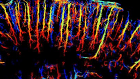
Image of the brain blood vessels, showing the density of the microbubbles. ESPCI/INSERM/CNRS
The ultrasonic waves can penetrate deep within tissue, but traditionally the resolution derived from this technique suffers from limitations. Due to the problem of diffraction, there is a trade-off between this resolution and how deep in the tissue the object being photographed is. Normally, doctors have to spend a long time to take a single high resolution image, but to get around this the researchers instead took lots of low resolution pictures, and then compared them all to build up an incredibly detailed snapshot.
With an ultrafast frame rate – in the region of 500 frames per second – the team accumulated over 75,000 images documenting two and a half minutes of the rat’s blood vessel network. They then took each image and subtracted the previous one allowing them to see each individual bubble. This gave them not only incredibly sharp pictures of the blood network with pixels 10 micrometers (0.01mm) in size, but also let them track the movement of the bubbles, and thus the flow of blood through the vessels and the direction it is moving in.
The researchers in France are not the only group working on how to build on ultrasound imaging techniques and perfect them to get higher and higher quality images, with another group in London and a third in Germany. They hope that with so many working on the problem, we could see the technique being used by doctors to track the flow of blood in and around tumors as they grow, allowing them to better understand how they might develop.