


Stanford scientists have used a combination of cutting-edge brain manipulation and monitoring techniques to identify a neural network that is associated with social interaction in mammals. By using light to stimulate specific neuronal populations within this circuit, the researchers were able to increase social interactions in mice. Conversely, when they inhibited the activity of this circuit the mice were less inclined to interact with others. The study has been published in Cell.
While previous studies have shed light on the systems implicated in social behavior at the level of the neurotransmitters involved, little was known about the neuronal circuits involved in social interactions. In order to further our knowledge of this subject, Stanford scientists headed by Karl Deisseroth employed a technique called optogenetics to both manipulate and monitor specific populations of brain cells in mice whilst observing the apparent effects on the behavior of the animals.
Deisseroth actually pioneered this neuromodulation technique which involves expressing light-sensitive proteins in neurons and then inserting an ultra-thin optical fiber near the brain region in question. Scientists can then use light to stimulate or inhibit these photosensitive cells whilst recording their activity. Since the animal is alive during the entire procedure, the researchers are able to investigate how manipulating certain cells affects behavior.
Deisseroth’s team used optogenetics to examine the relationship between social behavior in mice and an area of the brain called the ventral tegmental area, which is located in the brain stem. This region is heavily involved in the reward pathway, producing pleasure responses when an animal engages in certain activities such as eating or mating.
The VTA communicates with other brain regions via projections of neural fibers that release various neurotransmitters, including dopamine. While abnormal activity in the VTA has been associated with drug abuse and depression, little was known about its involvement in social activity.
The researchers chose to use only female mice in their experiments to make sure that mating- or aggression-related behaviors could not be confused with social behaviors. The team expressed photosensitive proteins in dopamine-secreting (dopaminergic) neurons within the VTA and then either selectively stimulated or inhibited these cells using light.
They found that enhancing the activity of these cells made the mice more inclined to engage in social behaviors with other mice. When a new mouse was introduced into the cage of a VTA stimulated mouse, the host would approach the newcomer and sniff it. However, when they inhibited the neurons, the mouse showed less interest towards the guest. Furthermore, they found that the effects were specific for social interaction as manipulating these neurons did not affect interactions with novel objects such as balls.
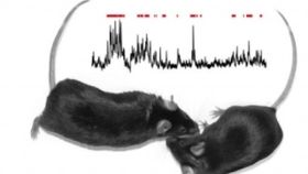
The researchers wanted to take this one step further by discerning which dopaminergic projections from the VTA were involved in carrying the signals that encourage social interaction. To do this they invented a novel monitoring technique, dubbed fiber photometry, which enabled the scientists to distinguish small signals from the brain’s background noise. The method involved measuring calcium flux, or the changes in calcium ion concentrations, which accompanies the signaling activities of neurons.
By combining this technique with optogenetics, the researchers found that a particular projection connecting the VTA with a mid-brain structure called the nucleus accumbens encodes and predicts certain features of social interaction.
Finally, the researchers manipulated dopaminergic cells within the nucleus accumbens and investigated how this affected the social exploration of the mice. They did this by replacing a type of dopamine receptor known as D1 with a light-inducible version. By stimulating these D1 cells in the nucleus accumbens, the researchers were once again able to enhance social exploration in the mice. Furthermore, they were able to verify these results by inhibiting these cells with a specific drug that blocks the activity of D1 receptors which reversed the effects.
“Every behavior presumably arises from a pattern of activity in the brain, and every behavioral malfunction arises from malfunctioning circuitry,” said Deisseroth in a news-release. “The ability, for the first time, to pinpoint a particular nerve-cell projection involved in the social behavior of a living, moving animal will greatly enhance our ability to understand how social behavior operates, and how it can go wrong.”