


A collaboration of researchers from MIT and the University of Vienna managed to map a worm’s nervous system as the action happens. The results of the study have been published in the journal Nature Methods.
Neurological activity, on its most basic level, involves the passing of calcium ions from one cell to another. When fluorescent markers bind to these ions, researchers can tell where the action is occurring. However, those methods typically are only able to see a small section of activity, and lack the bigger picture. The researchers developed a method to view these biomarkers in 3D at the millisecond timescale across the whole brain. This real-time imagery gives an unprecedented amount of detail to understand the full scope of neuronal activity as different parts work together to perform a task.
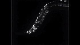
The researchers chose to study the brain of Caenorhabditis elegans, which is a simple worm with only 302 neurons. Humans, as a comparison, are estimated to have about 100 billion neurons with 100 trillion synapses. The 3D map of the C. elegans’ brain was created using a modified light-field microscope. This is the first time that equipment has been used for this kind of application. Instead of looking at each biomarker individually with one beam of light, the light is refracted into about 400 different parts, which a computer then puts together to create the map.
“Looking at the activity of just one neuron in the brain doesn’t tell you how that information is being computed; for that, you need to know what upstream neurons are doing. And to understand what the activity of a given neuron means, you have to be able to see what downstream neurons are doing,” lead researcher Edward Boyden of MIT said in a press release. “In short, if you want to understand how information is being integrated from sensation all the way to action, you have to see the entire brain.”
“We don’t really know, for any brain disorder, the exact set of cells involved,” he continued. “The ability to survey activity throughout a nervous system may help pinpoint the cells or networks that are involved with a brain disorder, leading to new ideas for therapies.”
The researchers are moving forward with this approach, trying to increase resolution. While they are able to scan markers more quickly than traditional methods, the resolution has suffered in the process and certain aspects get blurred. Increased computing power will also be needed to generate clearer pictures more quickly, as one second of imaging data can take several minutes to process and analyze. They are also seeking to blend this approach with optogenetics, which uses modified light-sensitive cells to regulate activity. This could bring resolution so advanced that researchers would be able to see individual dendrites on individual neurons as they pass the calcium ions along.