

Following five years of technological tinkering, researchers at the European Molecular Biology Laboratory (EMBL) have captured real-time images of specialized cells munching on neural connections in a mouse brain.
The group’s findings, reported in Nature Communications, further illuminate the physical process – called pruning – that streamlines the maturing mammalian brain by eliminating the excess neural connections that form during frenzied early development.
Neuroscientists have recently confirmed that the cell type behind this brain bonsai-ing are microglia – a type of immune cell that circulates through central nervous system tissue looking for things that don’t belong. When they come across cellular debris or pathogens, microglia remove the object though phagocytosis (i.e., they eat it).
Naturally, it was theorized that microglia use the same consumption capabilities to prune synapses, yet until now, no one had ever seen it happen.
“To the best of our knowledge,” the EMBL authors wrote, “our data are the first time-lapse images to directly demonstrate the active engulfment of synaptic material by microglia.”
However, the images revealed that microglia act much more delicately than ever expected. Rather than biting off entire synapses, microglia appear to only trim off small portions of the presynaptic axon, the area that stores neurotransmitter molecules.
Such restrained eating is referred to as trogocytosis, from the Greek word for “nibble”.

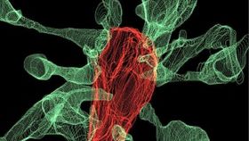
Multiple synapse heads send out filopodia (green) converging on one microglia (red). Image courtesy of L. Weinhard, EMBL Rome
Perhaps most surprisingly, the authors also saw that the presence of microglia in the brain’s memory center, the hippocampus, can cause postsynaptic dendrites to shoot many small outward projections called filopodia, creating new connections instead of destroying them. In one striking case, 15 synapse heads sent out projections toward a microglia as it nibbled on a synapse, suggesting these cells not only eliminate synapses but also induce growth and rearrangement.
"This shows that microglia are broadly involved in structural plasticity and might induce the rearrangement of synapses, a mechanism underlying learning and memory," said first author Laetitia Weinhard in a statement.
Obtaining these revolutionary images required the EMBL team to combine two microscopy systems: light sheet fluorescence microscopy, a laser beam-based method for imaging sub-cellular scale slices of organic material without destroying the sample, and correlative light-electron microscopy, a technique that combines an electron microscope with a light microscope to view cellular scale objects at high resolution.
According to lead researcher Cornelius Gross, the group’s subsequent investigations will focus on the role of microglia in the development of schizophrenia and depression – two disorders that are closely tied to irregularities in the hippocampus.
"This is what neuroscientists fantasized about for years, but nobody had ever seen before," said Gross. "These findings allow us to propose a mechanism for the role of microglia in the remodeling and evolution of brain circuits during development."